Macquiarie Neuro Surgery & Spine
    • About
      Our Team

Chiari Malformation


Some patients with Chiari malformation have no symptoms. For others, the condition may lead to serious complications.

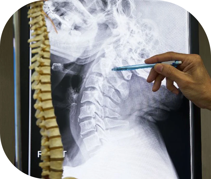
Patients with Chiari malformation require management by an experienced neurosurgical team. Specialists at Macquarie Neurosurgery & Spine are some of Australia’s leading experts on this condition and its complications. 

What is Chiari malformation?

Chiari malformation is a structural abnormality that forms during foetal development. The cerebellum is the back part of your brain, and it coordinates balance and movement. Normally, the cerebellum is found within your skull. In patients with Chiari malformation, part of the cerebellum is squeezed out into the upper spinal canal.

Chiari malformation can block the normal flow of cerebrospinal fluid (CSF) between the brain and spinal cord, causing increased pressure on these structures.

There are four different types of this condition. People with Chiari malformation type I may or may not experience symptoms. Type II Chiari malformation (historically known as Arnold-Chiari malformation) is generally more severe and is often associated with myelomeningocele (a type of spina bifida that causes paralysis in the legs). Types III and IV are rare. When they do occur, they cause severe problems from infancy and are often associated with other serious congenital defects.

Where does the name come from?

Chiari malformations were first described in 1891 by an Austrian pathologist named Hans Chiari (1851-1914).

What are the symptoms of Chiari malformation?

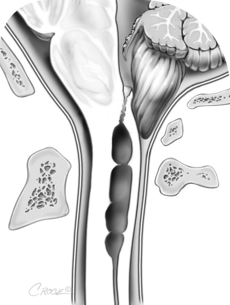
Chiari malformation is closely linked with syringomyelia.

Syringomyelia is the formation of cysts in the spinal cord. It can cause a wide range of symptoms, but typically leads to muscle weakness or sensation loss in the upper body, arms, and hands.

Other Chiari malformation symptoms could include:

  • Severe head and neck pain (often made worse by straining, sneezing, or coughing)
  • Dizziness or balance problems
  • Spasticity (abnormally stiff muscles)
  • Visual disturbances (like double or blurred vision, or hypersensitivity to bright lights)
  • Sleep apnoea
  • Cognitive impairment (also called “brain fog”).

Some patients with Chiari malformation also have spinal deformities like scoliosis. Patients with type II Chiari may develop hydrocephalus, a serious condition caused by excess CSF within the ventricles (cavities) of the brain.

Not all patients with Chiari malformation develop symptoms. Quite frequently, the condition is found incidentally when brain imaging is done for other reasons.

Why does Chiari malformation occur and who does it affect?

Chiari malformation can occur during pregnancy or later in life.

Primary or congenital Chiari malformation

This is the most common type of Chiari malformation and occurs during foetal development.

Many factors can affect foetal development, potentially leading to Chiari malformation. They include:

  • Genetic mutations
  • Lack of necessary vitamins and nutrients in pregnancy (like folic acid)
  • Infection or a high fever during pregnancy
  • Exposure to hazardous chemicals, illegal drugs or alcohol.

Secondary or acquired Chiari malformation

Chiari malformation can also happen later in life. It can happen when too much cerebrospinal fluid is drained from the spine after an injury, disease or infection.

How is it diagnosed?

Chiari malformation may be diagnosed:

  • Incidentally when the patient is being tested for other conditions
  • On ultrasound images before birth (in some cases)
  • After the diagnosis of other congenital conditions like spina bifida, which are known to be associated with Chiari malformation.

Diagnosing Chiari malformation may involve:

  • A physical examination to assess balance, touch, reflexes, sensation and motor skills
  • An assessment of cognitive function and memory
  • Imaging tests to show Chiari malformation, bone abnormalities that may be associated with it, or hydrocephalus.

Imaging tests for Chiari malformation

Advanced imaging tests enable us to diagnose Chiari malformation and determine its cause. These precise diagnostic methods are vital to the identification of Chiari malformation.

Magnetic resonance imaging (MRI) produces detailed 3D images of the brain structure. Both conventional and phase contrast MRI sequences can be used to assess the degree of hindbrain migration.

A computerised tomography (CT) scan may also be used. This uses X-rays to obtain cross-sectional images of the brain.

At Macquarie, we use advanced imaging such as dynamic MRI to gain a better understanding of the severity of a Chiari malformation and to guide treatment.

How is Chiari malformation treated?

Treatment of Chiari malformation (and associated syringomyelia) is dependent on the type of malformation and its symptoms.

If the malformation is not causing any symptoms, and has not led to syringomyelia, treatment may not be necessary. If there are symptoms, surgery may be considered to relieve pressure on the spinal cord and lower part of the brain, and to restore normal CSF flow.

SymptomsTreatment
None – and no syringomyeliaNone needed
Headaches – but no syringomyeliaPain relief
Persistent, unrelieved headaches, other symptoms or abnormal findings on neurological exam Surgery and ongoing management

Chiari decompression surgery

Chiari decompression surgery is a type of craniotomy that involves removing a small part of the base of the skull, as well as the back part of the first few neck vertebrae. A tissue graft (usually taken from the patient) may be used to further widen the canal.

This surgery is done to:

  • Make more room for the herniated brainstem (cerebellum)
  • Relieve pressure on the brain
  • Help to restore the normal flow of CSF around the brain – which may improve any associated conditions such as hydrocephalus or syringomyelia.

Potential complications of surgery

All operations carry a risk of haemorrhage or infection. Although rare and usually able to be managed, there is always a chance that haemorrhage or infection can be serious.

Particular complications for Chiari surgery include:

  • Meningitis
  • CSF leak of pseudomeningocele
  • Hydrocephalus
  • Tethering of the graft to the brain stem or cerebellum
  • Failure of a syrinx to resolve.

In some cases, further surgery may be necessary.

View a video by Professor Stoodley on this topic:

Play Video

Our expertise in Chiari malformation

Research

Macquarie Neurosurgery and Spine’s research over 20 years has contributed to advances in the management of Chiari malformation and syringomyelia. Our neurosurgeons maintain up-to-date knowledge and skills in supporting patients with Chiari malformation. We are committed to evidence-based care that optimises patients’ health outcomes.

Our current NHMRC-funded research is focused on understanding how Chiari malformations cause headache and cognitive impairment and how syringomyelia develops. We are aiming to determine the technical goals of surgery, which are currently unclear. Our research is also supported by the Column of Hope.

You can arrange a consultation with us at one of our NSW or ACT locations, by calling us on 02 9812 3900. Alternatively, you can request an appointment via our enquiry form.

Disclaimer

All information is general and is not intended to be a substitute for professional medical advice. Macquarie Neurosurgery and Spine can consult with you to confirm if a particular procedure is right for you.

References